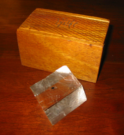
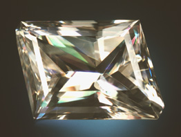
 Optical quality Iceland spar calcite with box carved by Leif Karlsen. Rhomb 65x45x35 mm Photo: E. Skalwold |
Historically a Mineral of Profound Importance For a historical perspective on Icelandic Spar calcite and its impact on science, please see the delightful paper written by Dr. Leo Kristjansson on "Iceland Spar: The Helgustadir Calcite Locality and its Influence on the Development of Science" (pdf file, 559 KB reproduced with permission of the author). A more recent and vastly updated paper in Icelandic will be available soon in English. |
| Update 12/26/15: For an in-depth look at the optical properties of calcite which makes it suitable as the Viking Sunstone, see Skalwold, E.A. and W.A. Bassett. (2015) Double Trouble: Navigating Birefringence. Chantilly, VA: Mineralogical Society of America. 20 pages. ISBN 978-0-939950-02-7 (now available from the MSA as premium quality hardcopy booklets here or as PDFs here). | 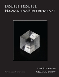 |
| Shown at right: parallel growth of two prismatic crystals, one of them dominant and partially doubly terminated. This specimen was obtained from Jordi Fabre, Fabre Minerals, and is much prized for its clarity and relatively rare prismatic form (larger image). The cleavage planes are clearly visible; dropping these prisms to the floor would sadly result in many much smaller, but identical cleavage rhombs, such as the one shown above and in the drawing below. Also see: optical quality calcite. 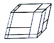 |
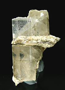 Calcite Prisms, Second Sovietsky mine, Dalnjegorsk, Primorskiy Kray Russia 6.8 × 4 × 3.1 cm Photo courtesy of Jordi Fabre, Fabre Minerals. |
| Calcite has three directions of perfect cleavage which reflect its three-fold symmetry. Each cleavage direction is inclined at the same angle to the c-axis; none are perpendicular or parallel to the c-axis (in calcite this is also the optic axis direction). When a crystal is broken, it tends to break on these directions forming a rhombohedron or "rhomb" - this is what was employed by Karlsen in his theory of the Viking Sunstone. These could be found in Iceland on the surface scree. | |
| The stats: Trigonal crystal system / Uniaxial negative / Refractive Index: ω= 1.658, ε=1.486 (sodium light) birefringence = 0.172. What does all that mean? Well, when light enters the calcite crystal it slows down and at some angles in which it strikes the surface (angle of incidence), it breaks into two light rays named ω and ε. Right after the interface of the air and crystal surface, the two rays bend at different angles and travel at different speeds through the calcite. They also become what is known as plane polarized. When they leave the crystal they become parallel. You don't have to know all that - it just explains why you see double through a crystal in some directions (see below). So if a speck of dirt or dot of tar is on one side of cleavage rhomb, you will see two dots looking from the other side - or if you place the rhomb over words on a paper, you will see them doubled. Those two refractive indices listed above represent the slowing of the two rays of light travelling through the crystal. The greater the difference between them, the more the doubling will be observed at particular orientations. (note: while the birefringence is greatest perpendicular to the optic axis, the two rays which are travelling at different speeds are not bending so there is no doubling observed -see figure 1 below - the unaided eye does not discern the two rays.). A crystal of quartz has a much lower difference or birefringence than calcite. A peice of quartz and a piece of calcite of exactly the same size and orientation will have a much different spread of images - the quartz will hardly be noticable unless it is a very, very thick peice while the calcite of the same size will show noticable doubling. | |
| An aside for the those interested in gems: Considered a collector gemstone because of its low hardness, calcite is not suitable for wear except with extreme care. However, a faceted optical quality calcite can be quite beautiful. Pockets of Iceland spar were found in the late 1960s in New York State which yielded rhombohedral crystals over 60 cm across. Master faceter Arthur Grant cut some of these which later became part of the gem collection of the Smithsonian Institution in Washington, D.C. as well as several other major museums. In the Winter 1984 issue of Gems & Gemology, Hurlbut and Francis describe the optics resulting from the twinning commonly found in these stones. A fantastic kaleidoscope of colors occurs as the light is broken up into progressively more pairs of rays as it crosses the twinning planes within the stones (Robinson, G.W. and Chamberlain, S.C., 2007). |
 St. Joe Minerals mine Balmat, St. Lawerence County, New York Photo courtesy of G. Robinson. |
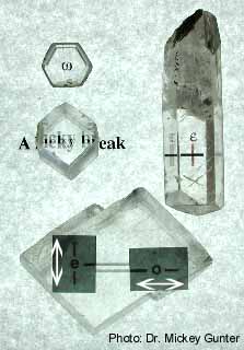 Calcite Crystals and Cleavage Rhombs Photo courtesy of Dr. Mickey Gunter. Gunter, M.E. (2003) "A lucky break for polarization: The optical properties of calcite." In extraLapis, Calcite, 4, 40-45, East Hampton, CT. Figure 1 |
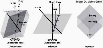 Refraction in Calcite Rhomb Image courtesy of Dr. Mickey Gunter. Dyar, M.D. and Gunter, M.E. (2007) Mineralogy and Optical Mineralogy. Mineralogical Society of America, Chantilly, Virginia, 708 pp. Figure 2 |
| Figure 1 shows calcite crystals and cleavage rhombs demonstrating the visual effects of light propagating through them. The top left crystal is the basal form and is shaped like a hexagon. Light behaves as the ordinary ray ω and travels along the c-axis straight out of the screen at you. The light remains unpolarized, does not break into two rays and therefore you don't see a double image. In the long prism form on the right, the c-axis is parallel to your screen and light is passing through the crystal perpendicular to the c-axis - that is straight out of your screen at you again. Even though the light travels along two seperate vibration directions in this orientation, the classic doubling effect does not occur. In reality the light entering the crystal becomes two rays traveling at different speeds. The single image below the crystal becomes two which are aligned one on top of the other - appearing as a single image. Because of its perfect cleavage, calcite is most often found as a rhombohedron. Light entering the crystal perpendicular to the face of the crystal ("normal") is neither parallel nor perpendicular to the optic axis as described in the crystals above. In the rhomb doubling occurs; that is, two images are produced as described below. The middle crystal shows a rhomb (rhombehedron) which has had the edges cut off on the left and right. The text on the paper underneath appears double when viewed through the rhomb. What you are observing is double refraction in which light entering the crystal is slowed and split into two mutually perpendicular plane polarized rays moving at different speeds: the ordinary ray (o-ray or ω) and the extraordinary ray (e-ray or ε). The intact rhomb below it shows the same effect, although this time 2 sheets of polarizing filter are placed on top of the rhomb with their transmission directions indicated by the white arrows. Notice that the images of the line underneath is seen doubled between the sheets, but singular through the sheets. The left sheet reveals the image generated by the e-ray; the right sheet reveals the image generated by the o-ray. This effect can also be observed by inking a dot on a peice of paper and placing a calcite rhomb over it. One will observe two dots from above. If the rhomb is rotated while kept flat on the paper, the dot produced by the e-ray will rotate around the stationary dot transmitted by the o-ray. The thicker the rhomb, the more spread out the images of the dots will appear. Figure 2 details the passage of the rays through a calcite rhomb. The rhomb is shown in various orientations with the vibration direction of each ray indicated by cross-bars mutually perpendicular to each other on the respective rays. In the middle side view, the o-ray vibration indicators are perpendicular to your screen and the e-ray indicators are parallel to your screen. "WN" indicates "wave normal." | |
| Update 12/26/15: For an in-depth look at the optical properties of calcite which makes it suitable as the Viking Sunstone, see Skalwold, E.A. and W.A. Bassett. (2015) Double Trouble: Navigating Birefringence. Chantilly, VA: Mineralogical Society of America. 20 pages. ISBN 978-0-939950-02-7 (now available from the MSA as premium quality hardcopy booklets here or as PDFs here). | |
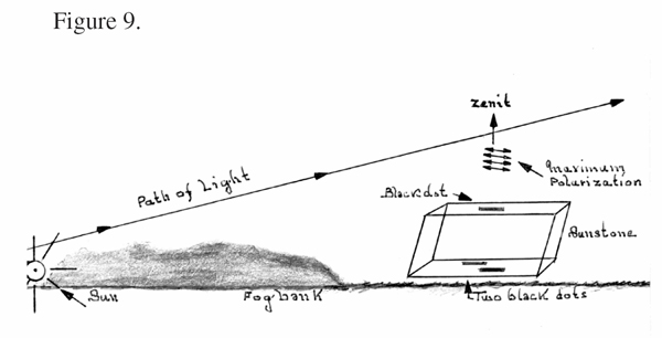
| Leif K. Karlsen's approach: | |
 Click to Enlarge |
 Click to Enlarge |
Return to the Viking Sunstone Main Page | |